Latest Headlines:

Archive: 03 Dec 2016


Sol Journal – December 3rd

December 3, 2016 | Permalink

It is all about science

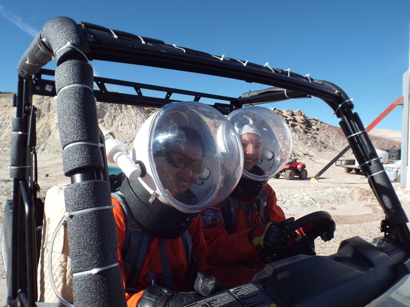
I’m not a scientist, but my favorite spot at Mars Desert Research Station is the science laboratory. It was build this summer and sometimes you can still sense little bit of paint smell. The dome has the biggest window in rectangular form, which opens the view to unique Utah desert landscape. This is the place where magic happens – the science discoveries and failures. Mars 160 mission has the main slant in field science research. Shannon Rupert, our Principal Investigator and backup crew biologist, along with members of our Earth-based science team from NASA’s Ames Research Center and Canadian Museum of Nature designed three science  projects for our crew astrobiologist/microbiologist Anushree Srivastava. She works closely with crew geologist Jon Clarke. Together they represent the geo-microbiology, which is the study of living and fossilized organisms in interaction with rocks. In order to lightened the heavy science research weight on Jon’s and Anushree’s shoulders, several of us were cross trained for the sampling and work in the laboratory. For me this was great opportunity to dive into microbiology world, even though I’m so far from understanding of it.

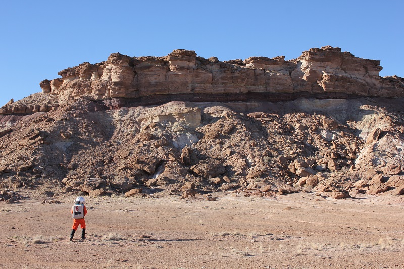
The first research is the mapping of biodiversity and distribution of lichens in this Martian analogue environment. Lichens are composite organisms made of fungi and algae, which usually form a colony. They can be found on the rocks and woody surfaces. If you see on a rocks a different colored patterns that might be lichen colonies. Why we are interested in studying lichens in this Martian analogue environment is because they can sustain in extreme conditions. Previous studies on lichens have demonstrated that they can survive in high radiation and desiccation. On Mars the radiation exposure on the surface is 30 μSv per hour during solar minimum. By studying the maximum constrains of where lichens can live, we can understand if they could survive in space environment. Additionally, they are useful to study the diversity of extreme life on Earth.

The second is the study of hypoliths, which are photosynthetic organisms that lives underneath translucent rocks in climatically extreme places, just like outside our station. Rock protects hypoliths from harsh ultraviolet radiation, dessication, and extreme temperatures. The rocks are generally translucent which allow hypoliths to receive light and moisture from the soil underside. There is a big possibility to find microorganisms living underneath the Martian rocks. Hypoliths will help humans to understand how to investigate microbial responses to environmental stressors.

The third study is finding traces of microlife trapped in ancient evaporites such as gypsum. Those microorganisms are halophiles. Halophiles (in Greek word for “salt-loving”) are organisms that thrive in high salt concentrations. They can be found anywhere with a concentration of salt five times greater than the salt concentration of the ocean. On Mars are plenty of gypsum deposits and the chances of finding microorganisms in it are high.

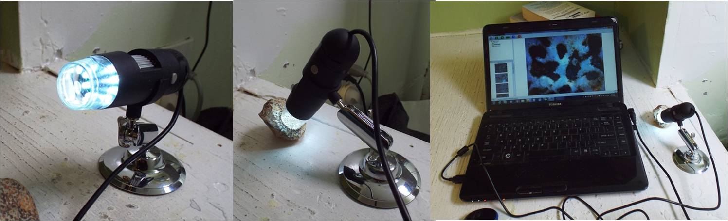
I just came back from science laboratory, where the magic was happening. Today Anushree did the plating to grow halophiles from soil samples collected during the EVA’s. But tomorrow will start the routine, observing everyday if there is any growth of microorganisms. It might take 15 days or months, but to be a scientist you have to be patient. And even more patient in the future when humans step on the red planet and the search for life on Mars or its traces starts.


Crew Photos – December 3rd

December 3, 2016 | Permalink

Colourful geology

Colourful geology


Jon scouting

Jon scouting


Lichens in the science dome

Lichens in the science dome


Phobos crew

Phobos crew


Anastasiya and mesa

Anastasiya and mesa


Anastasiya filming

Anastasiya filming


Anushree & Anastasiya in phobos & deimos

Anushree & Anastasiya in phobos & deimos


Picture of the Day – December 3rd

December 3, 2016 | Permalink

Phobos outside hab

Phobos outside hab


Sol Summary – December 3rd

December 3, 2016 | Permalink

Sol Summary Report (SSR):

Sol#70

Person filling out report: Annalea Beattie

Summary Title: O Happy Solar Day!

Mission Status on track:

Sol Activity Summary:

So this morning as I began to write to you, I stopped for a minute and thought about where I was. I’m in the Utah desert which is a really ancient amazing desert, in the Mars Desert Research Station, which is our home and a really great resource for science, and I’m sitting with the crew family at breakfast.

Perfect sun comes in through round windows of the hab, lots of chat about astronaut training and funding for the ExoMars Rover, the discussions about today’s EVA and who will cook lunch. These breakfast briefings are usually collegial and warm. We have a laugh and a joke, make each other coffee, check to see how we all slept and so on. Someone sits in the very sunny spot at the table. If it is Jon, he wears enormous black glasses. If it is Yusuke, he sits with his eyes shut, enjoying the warmth. Our crew always eats together. It’s a special time for us to relax and talk.

The most exciting news of today is that our solar panels are connected and now we have power in the laboratory. The lab has become an important working space for us all and that special window with the lovely view of the desert is spectacular.

Now with solar power, we can use all the equipment in the science dome.

Today Claude- Michel and Yusuke calibrated the pH sensors of the gardens and the temperature sensors of each spot. Then they wrote reports. I interviewed Yusuke again for space.com (there is a lot to say about him and his dome project), I wrote, made a spinach and cheese pie and baked some potatoes.

Out on the EVA, Anu drove the electric rover which was a big first for her! Alex’s suit interface was upset by the vibration on the ATV (there is no other explanation) and he had to come home. Jon and Anastasiya did some geological scouting with Anastasiya also filming messages for school kids. She is cleaning now.

What a whirlwind that woman is. Next dinner and early night.

Reports Submitted to CapCom:

  1.   Sol Summary- Annalea
  2.   Science  Narrative – Jon
  3.   Purple Mezuna Report-Yusuke
  4.   Anomaly Report – Alex
  5.  Pictures – Anastasiya
  6.   Photo of the Day – yes

MDRS Lessons of the Day

Alex learnt three letters of Russian.

Plans for tomorrow: Day off

Crew Physical Status: Yes, we are in excellent health!

Weather: still cold

Anomalies: no


Science Narrative – December 3rd

December 3, 2016 | Permalink

ROCKS UNDER THE MICROSCOPE

Jon Clarke

The study of rocks under the microscope is called petrography.  It is an essential tool in geology in its own right and as a precursor to more detailed studies.  Much petrography is done examining thin sections of rocks where they have been mounted on a glass slide and ground to a thickness of 30 microns (0.03 mm) and then examined with polarised light.  More specialised petrographic techniques include the study of polished sections with reflected light to study opaque minerals (minerography) and using transmission and scanning electron microscopy.

We do not have the facilities to make thin sections at MDRS, nor do we have polarising or electron microscopes, although an actual Mars mission might well do so.  But examining and documenting rocks with high resolution digital photography and under relatively low magnifying power reflected light microscopy is still a very useful tool.  Recent studies here at MDRS have shown this.

figure-1

Exotic clasts from the Fremont gravel terrace.  Left: siliceous sponge from the Permian Kaibab Limestone.  Fossil is 4.5 cm across.  Right: oolitic clast from the Triassic Sinbad Limestone.  Clast is 5 cm high.

Several days ago Anushree and I picked up some interesting geological specimens in the course of an EVA to sample lichens and hypoliths from gravel terraces.   These are shown in Figure 1, and consist of a Permian siliceous sponge, probably from the  Permian Kaibab Limestone and pebbles of oolitic limestone, probably from the Triassic Sinbad Limestone.  The nearest outcrop  upstream of these rock units are some 30-40 km away, in Capitol Reef National Park.

figure-2

Digital zoom of sponge fossil (left) and oolitic limestone (right). Light-coloured sponge skeleton contrasts with dark brown and yellow sediment fill of canals that are ~1mm across.  The ooids are the small spherical grains~0.5 mm in size.  These hand-held camera images have a resolution of ~20 microns per pixel, roughly equivalent to the MAHLI imager on the Curiosity rover on Mars.

Zooming in on both rocks with a high resolution digital camera we can see some detail, roughly equivalent to that captured by the MAHLI camera on the Curiosity rover and about four times better than the Microscopic Imager on the Mars Exploration Rovers.  The complex internal structure of the sponge is clearly visible.  The white material is chert, formed by alteration of the opaline silica spicules that originally made up the sponge’s skeleton.  The complex network of chambers through which the sponge pumped seawater and from which it filtered food particles, are visible by their dark brown or yellow sediment fills.  The close up of the oolitic limestone clearly shows the ooids, small round grains of calcium carbonate, that are diagnostic of warm, agitated clear-water conditions, super-saturated with calcium carbonate.  They are found today as vast shoals at depths of  between one and three metres deep in places like Great Salt Lake (Utah), the Bahamas, the Persian Gulf, and Shark Bay (Australia).

figure-3

Binocular microscope in the MDRS lab.

Looking at the rocks with the binocular microscope (X7 to X45 magnification) in our lab reveals many more details, including the presence of rare fragmentary microfossils in the limestone and a concentric and radial structure to the ooids.   The canal structure of the sponge is also revealed in more detail.  Unfortunately our binocular microscope, though very useful, cannot be used for photography.

figure-4

The USB microscope.  Left  – the microscope mount showing LED light sources surrounding the optics. Centre – the USB microscope positioned again it’s subject (the fossil sponge). Right – the USB microscope and plugged in showing user interface on laptop.

Fortunately Alex, our able commander, has brought on the expedition and extremely useful tool that allows us to examine rock surfaces at higher magnifications. This is the USB microscope, which comes with its own stand and LED light source and plugs into a laptop computer.  A small piece of software provides the user interface which includes image capture at a range of resolutions.    The USB microscope has a zoom lens from X10 to X200.

figure-5

USB microscope image of Permian sponge (left) and Triassic oolitic limestone (right). Image taken at X60 magnification.  Light-coloured cherty sponge skeleton contrasts with dark brown and yellow sediment fill of ~1 mm-sized canals.  The concentric structure of the ~0.5 mm diameter ooids is visible in this image.

The US microscope allows us to explore the rock surface in much greater detail, seeing details not visible in the digitally zoomed hand-held camera image. At X60 magnification, some further detail is visible along the margins of the canals in the sponge and the concentric structure of many of the ooids is evident.

figure-6

USB microscope images at X200 magnification.  Left: complex spicule structure visible inside white cherty skeleton of Permian sponge, microfossils are present in the dark brown and yellow sediment fill of the canals.  Right: Triassic oolitic limestone with both concentric and radial structure is visible in the ooids, along with the partial infill of inter-granular porosity by cement.

At even higher magnification (X200) some details of the complex spicule structure of the light coloured areas of the sponge are now visible, indicating that the sponge belongs to the Lithistid family.  A spiral-shaped silicified microfossil, probably a foraminifer, is visible in the sediment filling the canals, along with other, less identifiable fossil fragments.  The higher magnification has revealed that some ooids have a radial as well as a concentric fabric, and that the porosity between the ooids has been partly filled by bladed calcite cement.

figure-7

Traditional geological X10 hand lens

The beauty of the USB microscope is its simplicity, utility, and adaptability.  An instrument such as this is likely to find ready application of a crewed Mars mission.  Linked to a small tablet, or perhaps a screen inside a helmet, it would be an excellent substitute for that essential tool of the field scientist, the hand-held magnifying lens.  Though useful, such lenses are very difficult to use while wearing a space suit.  In addition to being of scientific utility, a tool such as the USB microscope can be used to detail with a range of engineering tasks which require inspection of small components or surfaces.  Such a tool would bridge the imaging gap between high resolution hand-held digital cameras and laboratory microscopes.